

What is a Mammogram?
Mammograms — a Critical Tool in Breast Health
Each year in the United States, hundreds of thousands of people are diagnosed with breast cancer. But early detection makes breast cancer easier to treat.
Mammograms can reveal breast health problems before there are any symptoms and can detect breast cancer in the earlier stages. Talk to your doctor about your breast cancer risk and schedule a mammogram today.
Our highly trained team of radiology experts use the latest digital mammography equipment, including 3D mammography, to perform more than 60,000 mammograms a year. Their experience — combined with the latest technology — means you get greater accuracy and peace of mind.
When Should You Have a Mammogram?
Most women should start screening mammograms at age 40. We recommend women aged 40-49 discuss their options with their primary care doctor and decide together based on the risks and benefits. All women over 50 should have mammograms at least every two years until they would not be healthy enough to undergo breast cancer treatment.
When you turn 40, ask your doctor for a breast cancer risk assessment and have your first mammogram no later than age 50. If you’re at higher risk due to family or personal history, your doctor may recommend that you begin regular screenings before 40.
After your first, you should schedule a mammogram at least every one to two years.
Schedule Your Mammogram at Baystate Health
People over 40 do not need a referral for a screening mammogram. You can call us or request an appointment online at one of our eight locations.
Learn More About Mammograms
What is a mammogram?
A mammogram is an X-ray of the breast. It can show potential problems — such as a breast lump, a cyst (a fluid-filled sac), or a build-up of calcium or fatty cells — before you or your doctor can feel them. It’s the only proven method for finding certain types of abnormal growths in the breasts.
There are two kinds of mammograms:
- Screening mammograms typically include two views of each breast taken from different angles. Five to 15 percent of people will need a follow-up exam or biopsy after a screening mammogram.
- Diagnostic mammograms are used to look for signs of breast cancer when you have symptoms – such as a lump, pain, or nipple discharge – or when your doctor sees suspicious results on a screening mammogram. Diagnostic mammograms are typically more detailed than screening mammograms.
You and your doctor will decide which type of mammogram is right for you.
Our advanced mammogram technology can detect trouble spots early and ensure you get the support and care you need as quickly as possible. The mammography technology we use includes traditional 2D (two dimensional) mammography and 3D (three dimensional) mammography (also called breast tomosynthesis or “tomo”), which uses the same X-rays as a traditional 2D mammogram but creates a 3D image of your breast from all sides.
Preparing for a Mammogram
When you make your appointment, you will receive preparation instructions, but there are some general guidelines.
- Before your mammogram, talk to your doctor about any new problems in your breasts.
- Tell your doctor about any surgeries you’ve had, hormone use, and family or personal history of breast cancer.
- Women should make their appointments for one week after their period. Your breasts are less likely to be tender at that time.
- If you have had a mammogram somewhere other than Baystate Health, bring those records to your appointment.
- Wear a top and a separate bottom, rather than a dress or one-piece outfit, so that you can undress only from the waist up.
- Caffeine can make your breasts tender, so avoid coffee, tea or caffeinated soft drinks for a few days before your mammogram.
- Don’t wear deodorant, perfume, lotion, or powder under your arms or on your breasts on the day of your appointment. These substances can cause your images to be less clear.
- If you have breast implants, be sure to share that when you make your appointment. You’ll need a technologist who is trained to X-ray patients with implants.
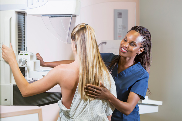
What to Expect During Your Mammogram
- You’ll be asked to remove your top and bra if you’re wearing one and put on a gown.
- You may sit in a waiting area before being taken to the room containing the mammography machine.
- You’ll be asked a few questions to confirm your identity and your health history.
- A specially trained technician will place one of your breasts onto the mammography machine platform.
- The tech will adjust the machine to gently compress (flatten) your breast.
- You may feel discomfort, but the exam shouldn’t be painful.
- Your technician will take two images of each breast: a top-to-bottom view and a side view. You’ll be asked to stand still for a few seconds while each picture is taken.
- Once they take pictures of one breast, they’ll do the same with the other.
- The entire exam should last about 20 minutes, with only a few seconds of compression while each picture is taken.
Compression is important because it:
- Evens out your breast thickness to help reveal any abnormal spots
- Helps lower the dose of radiation you’re exposed to
- Keeps your breast still so we can get more accurate, clearer pictures
Getting Your Mammogram Results
What is digital mammography?
Digital mammography is the standard in mammography technology. It produces clear, accurate X-rays in the form of digital files.
Digital mammograms have been shown to better detect breast cancer in people who are under age 50, have dense breast tissue, or are nearing menopause. They also make it easier to confidentially store and share mammogram results between breast surgeons and radiologists.
In addition to digital mammography, we use a sophisticated computer-aided design (CAD) program to help us interpret your mammogram. This software allows our radiologists to mark spots of potential concern, which can then be reviewed in follow-up exams.
When is 3D mammography used?
A 3D mammogram (also called breast tomosynthesis or "tomo") is a type of digital mammogram. It uses the same X-rays as standard 2D mammograms and it’s done in a similar way: your breast is compressed between two plates.
Unlike 2D mammograms, which take images only from the front and side, 3D mammograms take images, or "slices," of your breast from many different angles. This creates a 3D picture of your breast.
While 3D mammography may sound like an improvement over 2D mammography, it’s not right for every case. It can be useful for assessing the size of a cancerous growth and for screening breasts that are particularly dense.
3D mammograms are not covered by every insurance plan, and they are not necessary for every screening. We’ll discuss your options with you and decide if 3D or 2D is right for your situation.
Back to Top